The era of awake aware painless scarless endoscopic Spine surgeries is here
The spinal cord is the continuity of the brain and connects other parts of the body with the brain through the millions of nerves, taking information to and fro between the brain and other organs/parts of the body. The soft spinal cord is protected by the bony vertebral column of the back and in between the hard vertebral bones of the back, are the soft structures known as the intervertebral discs, which provide flexibility to the bony vertebral column, so that we are able to bend and twist our body.
Ageing leads to degenerative changes to the intervertebral disc structure, leading to tears and ruptures in the structure known as annulus fibrosus, which surrounds and encases an inner soft jelly-like substance known as the Nucleus Pulposus. The Nucleus pulposus also loses its strength and the disc can reduce in height and bulge outwards too. Once the degenerative changes occur in the disc, it leads to new abnormal nerves growing into the discs and now even normal movements of the neck, known as the cervical spine or the lower back known as the lumbar region, can lead to pain and stiffness in the neck or lower back after work or even prolonged sitting and bending forwards.

 Endoscopic spine surgery
The conditions which increase the probability of early degeneration of the intervertebral disc include genetics, smoking, a sedentary lifestyle, work involving vibrating machines, constant lifting of heavy weights and nowadays driving two-wheelers also contribute to neck pain and low back pain, also prolonged use of computers and laptops in improper positions, and using cell phones too much also contribute to this kind of neck pain and low back pain.
Sometimes when excessive and severe compressive and twisting forces are applied to the intervertebral disc, occurring in conditions like lifting heavy weights from the floor may result in complete total rupture of the intervertebral disc either due to a gradual weakening of the disc due to degeneration or due to sudden total failure of the integrity of the disc, the nucleus pulposus can rupture out of the disc breaking out of the encasing annulus fibrosus and prolapse into the spinal canal containing the spinal cord and compresses either the spinal cord or the nerve roots which come out of the spinal cord and go to the upper or lower limbs or to the other organs of the body like the urinary bladder and the bowels.
This causes the severe back pain or severe nerve root pain, which can radiate from neck to the hands or from the lower back to the leg and feet, commonly known as sciatica and it can cause numbness in the nerve distribution in the hand or leg and foot, and it can also lead to weakness of the muscles, the nerve supplies and controls and may lead to complete loss of control of the foot and ankle muscles known as foot drop and sometimes severe compression of the spinal cord can result in a condition known as Cauda Equina syndrome leading to loss of control of the urinary bladder or bowel control, situation which are to be dealt in an emergency fashion and require immediate surgery to remove the offending disc material.
Usually, if there is no major motor power weakness, it can be managed conservatively with medicines, treatment requires investigations like X-rays, some basic blood tests and an MRI and CT Scan needed to know the kind of damage that has occurred to the Spinal Column. Treatment include medicines to reduce pain and medicines to increase sensation and function of the nerves. Sometimes physiotherapy like cervical traction and IFT help to reduce arm pain or leg pain and help in the gradual reduction of pain over the course of two or three weeks.
If the pain is unremitting to conservative treatment or the severity of pain is increasing in spite of treatment and resulting in weakness of hand or foot or where there is loss of control of the urinary bladder or the bowel control, surgeons need to do surgery to remove the offending disc material compressing the thecal sac or the nerve roots within the spinal canal.
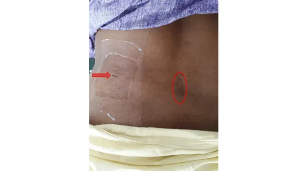 Traditional surgery scar in centre and endoscopic scar arrow mark in the side
Usually, the traditional surgery is done under general anaesthesia and it involves an incision in the midline of the back, which may vary in length between a minimum of 5 to even 10 cms, and the surgery may be done with the help of a microscope, involves cutting normal structures surrounding the vertebral column, like the back muscles, then involves removing some of the innocent by-standers and stabilizing structures of the vertebral column like the bony lamina and spinous processes, and other ligaments, which covers the spinal cord and finally after retracting the spinal cord and nerve root the offending disc can be removed and all this surgical procedure itself can sometimes result in unavoidable collateral damages to the normal muscles and bone and spinal cord and nerve root.
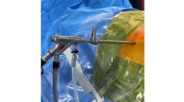
Some innovative and pioneering spine surgeons like Professor Pervez Kambin in the USA and Professor Hijikata in Japan in the 1980s and 1990s devised a surgery by going through the sides of the vertebral column by just dilatation of the muscles of the back. Dr Antony Yeung in Phoenix, Arizona, USA, developed special spinal endoscopes and other instruments to do proper disc excision surgery through just a 7 mm incision in the sides of the back, to remove the offending disc tissue with absolutely no collateral damage to the stabilizing normal structures of the vertebral column.
The greatest beauty of the innovation is that the surgery can be done under local anaesthesia by numbing a small area of skin and with the patient lying on his front comfortably fully awake and aware. There is very minimal blood loss and there is no need for blood transfusions too. For successful results, the surgery is done with advanced computer X-rays, and other special machines known as radio frequency cautery and lasers known as Holmium-YAG lasers which help to vaporize the disc or shrink the disc or abnormal bone growths which may come in the way of the surgery or which may also be compressing the thecal sac or the nerve root.
The greatest advantage of this Percutaneous Endoscopic spine surgery is that the patient can go home within 3 to 6 hours after surgery as a daycare procedure, and the patient’s post-operative rehabilitation is also very fast and quick and they can get back to normal activities within 2 to 3 weeks while in the traditional surgery, the patient usually requires around 3 to 5 days of hospital admission, have more pain for the first 3 weeks after surgery. Finally, there will be a big scar in the back also but with endoscopic spine surgery, with just an 8 mm incision in the sides of the back, the small scar just disappears like a small scratch in the back in the course of 3 months to 6 months, hence cosmetically very good for young patients, especially ladies. Naturally, worldwide this percutaneous endoscopic spine surgery has emerged as the gold standard surgery for spine disc problems in recent times.
Recent advances in the science of instruments and visualization technology of endoscopic instruments in the past 4 to 5 years have resulted in a complete revolution of spine care surgeries for old age people too. Now all kinds of spinal problems of aging due to wear and tear of the spinal column resulting in deformities of the spinal column and resultant compression to the spinal cord and nerve roots known as spinal canal stenosis, can now be corrected endoscopically.
While previous operations for such conditions were open surgeries which required long 15 to 20 cms of incisions, can now be done by endoscopic techniques involving just keyhole incisions of 1 cm, placed strategically in the back by properly trained spinal surgeons, can do this deformity correction or spinal canal decompression relieving the patients of their life-long sufferings without much post-operative sufferings for the patients. Such advanced instruments and surgeries are now available in Kauvery Hospital Tirunelveli and is a boon to patients and we can say that an new era of awake aware key hole endoscopic spine surgeries which are almost scarless and painless has come to the benefit of patients of the Southern tip of our motherland.
 Dr. K Sekar, MB., D.Ortho., Mch. Ortho, Consultant Orthopedic Spine Surgeon, Kauvery Hospital, Tirunelveli
Dr. K. Sekar, a highly experienced specialist in orthopaedics and spine surgery, is dedicated to providing expert care at Kauvery Hospital Tirunelveli. His extensive knowledge and skill in treating various orthopaedic and spinal conditions ensure that patients receive the highest quality of care and support throughout their treatment.
Disclaimer:
This content is sponsored and does not reflect the views or opinions of IE Online Media Services Pvt Ltd. No journalist is involved in creating sponsored material and it does not imply any endorsement whatsoever by the editorial team. IE Online Media Services takes no responsibility for the content that appears in sponsored articles and consequences thereof, directly, indirectly or in any manner. Viewer discretion is advised.
📣 For more lifestyle news, click here to join our WhatsApp Channel and also follow us on Instagram
Disclaimer: The copyright of this article belongs to the original author. Reposting this article is solely for the purpose of information dissemination and does not constitute any investment advice. If there is any infringement, please contact us immediately. We will make corrections or deletions as necessary. Thank you.
Read More

Consuming a plant-based diet does not necessarily mean it’s healthy; here’s why
While many view plant-based diets as a panacea for health, a new study published in The Lancet Regio...
Kusha Kapila feels ‘dehumanised’ after jokes made about her divorce and weight in roast video; how public shaming can impact individuals
Content creator-turned-actor Kusha Kapila has spoken out against her experience on comedian Ashish S...
Can having lemon juice and olive oil on an empty stomach relieve constipation?
Clearing your bowels in the morning is extremely important for overall wellness. But if you struggle...
Shweta Tripathi Sharma on how playing Golu Gupta in ‘Mirzapur’ has affected her personally: ‘It does take a toll, sometimes a lot’
The first time Shweta Tripathi Sharma went on stage was when she was only six years old. While she h...
We could not believe that pregnancy planning and hair wash have a connection, so we asked experts
There are various dos and don’ts when it comes to pregnancy planning: from eating certain foods, and...
Radhika Merchant wore rare natural Burma ruby jewellery set for her sangeet: Learn what makes it precious
Radhika Merchant, bride to her childhood sweetheart Anant Ambani, embodied timeless elegance at her ...